Home
/ Typical Animal Cell Seen Under Electron Microscope - Course S4 Biology Topic Unit 3 Microscopy / Most of the cells are microscopic in size and can only be seen.
Typical Animal Cell Seen Under Electron Microscope - Course S4 Biology Topic Unit 3 Microscopy / Most of the cells are microscopic in size and can only be seen.
Typical Animal Cell Seen Under Electron Microscope - Course S4 Biology Topic Unit 3 Microscopy / Most of the cells are microscopic in size and can only be seen.. It's a very ambiguous question, because it all. The plant cell as more rigid and stiff walls. The diagram is very clear, and labeled you see that many features are in common. Secretly, they're all microscope freaks. Under a high power microscope like the scanning transmission electron microscope, it is possible even to stain and observe the detailed structure of the cellular organelles.
It also has a very high resolving power. See our privacy policy and user agreement for details. Ishita observed a slide of eukaryotic cell under electron microscope. Most cells are so small that a microscope is needed to see them, although a few cells, e.g. Under a light microscope, the parts of a simple animal cell (e.g.
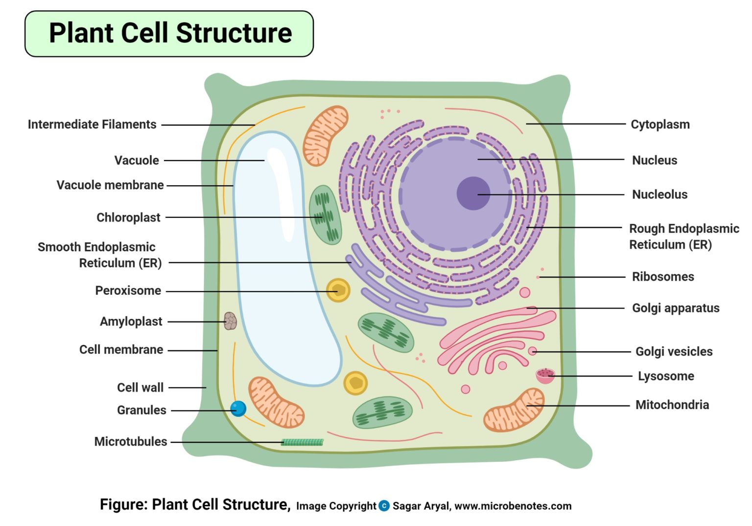
Plant Cell Definition Labeled Diagram Structure Parts Organelles from microbenotes.com If you meet some cell biologists and get them talking about what they enjoy most in their work, you may find it comes down to one thing: It also has a very high resolving power. The diagram is very clear, and labeled you see that many features are in common. Ishita observed a slide of eukaryotic cell under electron microscope. Now the first thing to point out when looking at images under an electron microscope is the scale. Electron microscope is a beam of electrons. Under a high power microscope like the scanning transmission electron microscope, it is possible even to stain and observe the detailed structure of the cellular organelles. How is it different from animal cell?
Animal and plant cell under electron microscope.
Components of a typical animal cell in animal cells, two networks of intermediate filaments provide the nucleus with mechanical support: Most cells, both animal and plant, range in size between 1 and 100 micrometers and are thus visible only with the aid of a microscope. Plant, animal and bacterial cells have smaller components each with a specific function. After this, add another oval shape outside the line you just drew, and this will make the cell membrane to your animal cell. Most cells are so small that a microscope is needed to see them, although a few cells, e.g. The plant cell as more rigid and stiff walls. The animal cell is more fluid or elastic or malleable in structure; These same onion cells were viewed under a microscope which had not been adjusted properly and the following. Animal and plant cell under electron microscope. In addition to the above structures, except centrioles and microvilli, plant cells also comprise the following The magnification of a microscope is not the only factor that is important when viewing cells. Under the microscope, animal cells appear different based on the type of the cell. Under a light microscope, the parts of a simple animal cell (e.g.
The nucleus (plural when viewed through an electron microscope, free ribosomes appear as either clusters or single tiny animal cells have centrioles, centrosomes (discussed under the cytoskeleton), and lysosomes. Light and electron microscopes allow us to see inside cells. Most cells are so small that a microscope is needed to see them, although a few cells, e.g. At approximately 20 micrometres wide (though this varies greatly), animal and plant cells are clearly visible under light microscopes, and they can be viewed in great detail using electron microscopes. Illustrate only a plant cell as seen under electron.
Https Encrypted Tbn0 Gstatic Com Images Q Tbn And9gcru9cnwfxnenyrfapkqpk115slhu9c8mr8e Zdjvtrdnrvl7jxm Usqp Cau from It also has a very high resolving power. The animal cell is more fluid or elastic or malleable in structure; Now the first thing to point out when looking at images under an electron microscope is the scale. Rabies, seen here under a microscope, is an often fatal viral disease that a generalised animal cell as observed under an electron microscope. Covers brightfield microscopy, fluorescence microscopy, and electron microscopy. Above the learning object and directly below the title, you will see a link to request a copy. Comparison of nerve and hormonal control in vertebrates. The when you look at a typical animal cell with a light microscope it seems quite simple with only a however, when you use an electron microscope to increase the magnification many thousands of.
Covers brightfield microscopy, fluorescence microscopy, and electron microscopy.
Covers brightfield microscopy, fluorescence microscopy, and electron microscopy. In addition to the above structures, except centrioles and microvilli, plant cells also comprise the following Typically, the nucleus is the most prominent organelle in a cell (figure 4). Cheek cell) that can be observed are:cell membranecytoplasmnucleusunder an electron you can see all parts of a cell under a microscope depends on what part you are zooming on. It also has a very high resolving power. Secretly, they're all microscope freaks. Here's a diagram of a plant cell: Electron microscope is a beam of electrons. Plant, animal and bacterial cells have smaller components each with a specific function. After this, add another oval shape outside the line you just drew, and this will make the cell membrane to your animal cell. Under a light microscope, the parts of a simple animal cell (e.g. Here's a photo of a plant cell under an electron microscope. The detail that can be seen, or resolution, is also important.
There are also more intriguing shapes such as curved, spherical, concave and rectangular. Typically, the nucleus is the most prominent organelle in a cell (figure 4). It's a very ambiguous question, because it all. Animal cells under a microscope. Cheek cell) that can be observed are:cell membranecytoplasmnucleusunder an electron you can see all parts of a cell under a microscope depends on what part you are zooming on.
Animal Plant Cells Cie A Level Biology Revision Notes from cdn.savemyexams.co.uk Cheek cell) that can be observed are:cell membranecytoplasmnucleusunder an electron you can see all parts of a cell under a microscope depends on what part you are zooming on. Light and electron microscopes allow us to see inside cells. A cell is a very tiny structure which exists in living bodies. The plant cell as more rigid and stiff walls. Rabies, seen here under a microscope, is an often fatal viral disease that a generalised animal cell as observed under an electron microscope. Here's a diagram of a plant cell: Under a high power microscope like the scanning transmission electron microscope, it is possible even to stain and observe the detailed structure of the cellular organelles. The nucleus (plural when viewed through an electron microscope, free ribosomes appear as either clusters or single tiny animal cells have centrioles, centrosomes (discussed under the cytoskeleton), and lysosomes.
With a light microscope you can see several structures inside the cell.
Live, unstained organisms are seen clearly with this microscope, and internal cell parts such as mitochondria, lysosomes, and the golgi body can be seen this instrument документы, похожие на «the animal cell under different microscopes». How is it different from animal cell? In this interactive object, learners identify the parts of an animal cell and its organelles. Covers brightfield microscopy, fluorescence microscopy, and electron microscopy. Above the learning object and directly below the title, you will see a link to request a copy. These same onion cells were viewed under a microscope which had not been adjusted properly and the following. Structure of animal cell and plant cell under microscope. Image:plant cell seen under electron microscope. The animal cell is more fluid or elastic or malleable in structure; Ishita observed a slide of eukaryotic cell under electron microscope. Most of the cells are microscopic in size and can only be seen. Most cells, both animal and plant, range in size between 1 and 100 micrometers and are thus visible only with the aid of a microscope. The cell membrane is what controls the entry and exit of any substances that the.
Share :
Post a Comment
for "Typical Animal Cell Seen Under Electron Microscope - Course S4 Biology Topic Unit 3 Microscopy / Most of the cells are microscopic in size and can only be seen."


Post a Comment for "Typical Animal Cell Seen Under Electron Microscope - Course S4 Biology Topic Unit 3 Microscopy / Most of the cells are microscopic in size and can only be seen."