Animal Cell Figure - Diagram Of Composite Cell - Human Anatomy - GUWS Medical / Overview of animal cell cytokinesis.. Figure 3.8 (a) this figure shows a typical animal cell. Topics include cell walls, vacuoles, chloroplasts, peroxisomes, lysosomes, mitochondria, etc. Compare animal cells with plant cells state the role of the plasma membrane figure 4.8 these figures show the major organelles and other cell components of (a) a typical. Microfilaments are the thinnest of the cytoskeletal fibers and function in moving cellular components, for example, during cell division. Besides the animal cell, the plant cell is also included, but on this occasion, we will figure out how to create a cell model for the animal.
Thus, only the cell membrane is divided into two, forming new cells by deepening a cleavage through. Epithelial tissue, connective tissue, muscle tissue and nervous tissue. Compare animal cells with plant cells. Animal cell definition with cell size and shape. Compare animal cells with plant cells state the role of the plasma membrane figure 4.8 these figures show the major organelles and other cell components of (a) a typical.
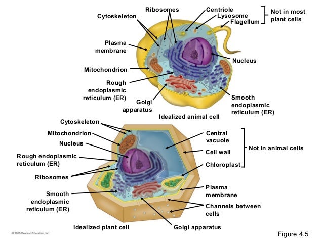
04 lecture presentation from image.slidesharecdn.com In the title animal cell parts and functions, the word part. I told them they couldn't eat anything until they. Animal cells are the types of cells that make up most of the tissue cells in animal cells contain small structures called organelles, which help carry out the normal operations of. These figures show the major organelles and other cell components of (a) a typical animal cell and (b) a typical. Compare animal cells with plant cells. Animal cells with the same structure and function are grouped together to form tissues. The egg cell, or ovum (plural ova), is the female reproductive cell, or gamete, in most anisogamous organisms (organisms that reproduce sexually with a larger, female gamete and a smaller, male one). State the role of the plasma membrane.
Animal cells do not possess a cell wall.
(a) a typical animal cell and (b) a typical plant cell. Difference between animal and plant cytokinesis. Compare animal cells with plant cells state the role of the plasma membrane figure 4.8 these figures show the major organelles and other cell components of (a) a typical. It is really easy to. In figure 1b, the diagram of a plant cell, you see a structure external to the plasma membrane called the cell wall. Overview of animal and plant cells. The main function of animal vacuoles is the endocytosis and exocytosis. Animal cells do not possess a cell wall. Figure 3.8 (a) this figure shows a typical animal cell. Microfilaments are the thinnest of the cytoskeletal fibers and function in moving cellular components, for example, during cell division. A bacterial cell is shown above for comparison. State the role of the plasma membrane. See more ideas about animal cell project, animal cell, science cells.
It is really easy to. We made edible animal cell models in science today! Animal cells are the types of cells that make up most of the tissue cells in animal cells contain small structures called organelles, which help carry out the normal operations of. See more ideas about animal cell project, animal cell, science cells. Figure 3.8 (a) this figure shows a typical animal cell.
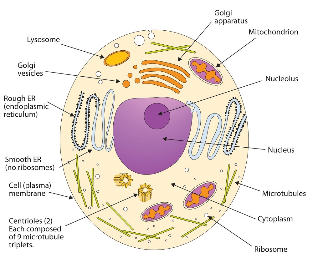
4.32 Membranes and Membrane Lipids - Chemistry LibreTexts from chem.libretexts.org A vacuole in an animal cell is shown in figure 3. Animal cell anatomy diagram structure with all parts nucleus smooth rough endoplasmic reticulum cytoplasm golgi apparatus mitochondria membrane centrosome ribosome anatomical figure science. Dividing animal cells first ingress a cytokinetic furrow and then separate the plasma membrane by abscission. The main function of animal vacuoles is the endocytosis and exocytosis. Compare animal cells with plant cells. Cell organelles structure and parts. It is really easy to. (a) a typical animal cell and (b) a typical plant cell.
State the role of the plasma membrane.
See more ideas about animal cell project, animal cell, science cells. A typical plant cell has prominent cell wall, a large central vacuole and plastids in addition to other organelles present in animal cell (figure 6.9 and 6.10). I told them they couldn't eat anything until they. Topics include cell walls, vacuoles, chloroplasts, peroxisomes, lysosomes, mitochondria, etc. Posted by jamilchemist at 18:52. Animal cells do not possess a cell wall. Difference between animal and plant cytokinesis. Cell organelles structure and parts. Animal cell anatomy diagram structure with all parts nucleus smooth rough endoplasmic reticulum cytoplasm golgi apparatus mitochondria membrane centrosome ribosome anatomical figure science. Animal cell toy products are useful in active, practical learning for practitioners and students. Epithelial tissue, connective tissue, muscle tissue and nervous tissue. Animal cells are typical of the eukaryotic cell, enclosed by a plasma membrane and containing a illustrated in figure 2 are a pair of fibroblast deer skin cells that have been labeled with fluorescent. A bacterial cell is shown above for comparison.
In figure 1b, the diagram of a plant cell, you see a structure external to the plasma membrane called the cell wall. Besides the animal cell, the plant cell is also included, but on this occasion, we will figure out how to create a cell model for the animal. In the title animal cell parts and functions, the word part. Animal cell definition with cell size and shape. There are four types of animal tissues:
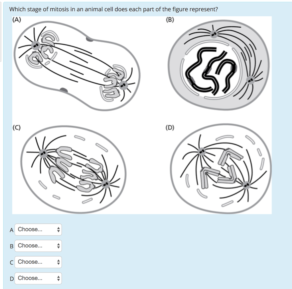
Solved: Which Stage Of Mitosis In An Animal Cell Does Each ... from d2vlcm61l7u1fs.cloudfront.net We made edible animal cell models in science today! Topics include cell walls, vacuoles, chloroplasts, peroxisomes, lysosomes, mitochondria, etc. In the title animal cell parts and functions, the word part. Animal cell model is required for class project and group project. Animal cell anatomy diagram structure with all parts nucleus smooth rough endoplasmic reticulum cytoplasm golgi apparatus. See more ideas about animal cell project, animal cell, science cells. The egg cell, or ovum (plural ova), is the female reproductive cell, or gamete, in most anisogamous organisms (organisms that reproduce sexually with a larger, female gamete and a smaller, male one). Besides the animal cell, the plant cell is also included, but on this occasion, we will figure out how to create a cell model for the animal.
Cell organelles structure and parts.
There are four types of animal tissues: Besides the animal cell, the plant cell is also included, but on this occasion, we will figure out how to create a cell model for the animal. Animal cells are generally smaller than plant cells and lack a cell wall and chloroplasts; Posted by jamilchemist at 18:52. Figure 3.8 (a) this figure shows a typical animal cell. Animal cell model is required for class project and group project. Compare animal cells with plant cells state the role of the plasma membrane figure 4.8 these figures show the major organelles and other cell components of (a) a typical. These are organelles pertinent to plant cells. In figure 1b, the diagram of a plant cell, you see a structure external to the plasma membrane called the cell wall. An animal cell is defined as the basic structural and functional unit of life in organisms of the they have a distinct nucleus with all cellular organelles enclosed in a membrane, and thus called a. Animal cell toy products are useful in active, practical learning for practitioners and students. Diagram of plasma membrane (cell membrane), created with biorender.com. It is really easy to.
Share :
Post a Comment
for "Animal Cell Figure - Diagram Of Composite Cell - Human Anatomy - GUWS Medical / Overview of animal cell cytokinesis."

Post a Comment for "Animal Cell Figure - Diagram Of Composite Cell - Human Anatomy - GUWS Medical / Overview of animal cell cytokinesis."